
If your dog has been diagnosed with a deep corneal ulcer and a grafting procedure has been recommended. The information below will help you to understand your options and what to expect following the operation.
Deep corneal ulcers
All corneal wounds are referred to as ‘corneal ulcers’. There are many reasons why a patient may develop a corneal ulcer – including trauma, infection, rubbing from in-turned eyelids and dry eye. Superficial corneal ulcers are painful but do not pose a threat to the integrity of the globe and are usually treated medically - with few exceptions.
Deep corneal ulcers however pose a threat as they may result in corneal perforation (rupture) – which is extremely painful and sight threatening and often require emergency surgical intervention. Surgeries for deep corneal ulcers usually involve placement of a ‘graft’ into the defect – which provides structural support to the eye. If a deep corneal ulcer, which puts the eye at risk of rupture, is diagnosed, then a pro-active approach with surgery is usually preferable.
This does not mean that deep ulcers cannot heal with medical treatment alone – but there is a significant risk that the eye could be lost if the ulcer ruptures under medical treatment. We can also operate on corneal ulcers that have already ruptured if the patient is presented quickly – but this surgery is more challenging and has a higher risk of complications that surgery carried out before the ulcer ruptures.
What surgical options to support a deep corneal ulcer are available?
There are many different grafting procedures available, but the two types of graft that we most commonly use here at EVC are the following:
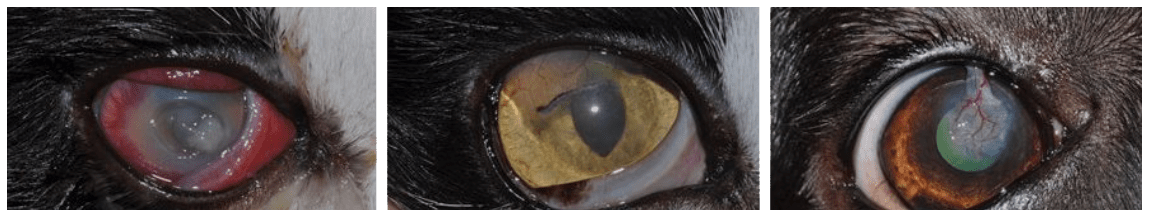
Conjunctival pedicle graft (also called ‘CPG’)
In this grafting procedure, a thin piece of the conjunctiva (the pink skin that covers the white of the eye) is sewn into the defect. The conjunctiva provides some structural support but also brings blood vessels into the ulcer – which in turn allow much faster healing of the wound than would usually be seen in the clear cornea, which is free of blood vessels. The conjunctival pedicle graft is especially suitable for small but deep corneal wounds and in cases of active corneal infection – as the blood supply of this type of graft helps to actively fight infection.
Corneo-conjunctival transposition graft (also called ‘CCT’)
In this grafting procedure, a partial-thickness piece of healthy corneal tissue adjacent to the wound is harvested together with some attached conjunctiva, mobilised and sutured into the defect. This graft is especially suitable for larger deep defects as it brings superior stability. In addition, this graft will give the best cosmetic and functional result as the piece of cornea sutured into the original defect usually will be clear again. In some cases of severe infectious corneal ulceration, we may however advice against a CCT as it could be prone to destruction by the infection. In both of the above grafting procedures, it is essential that the harvest and suturing of the graft is carried out under the operating microscope. The ophthalmologists at EVC have been especially trained in microsurgical skills which allow handling of the tissue and suture material (which is barely thicker than a hair!) with minimal trauma.
Find out more about our ophthalmology referrals
What do I expect after my pet has undergone a corneal grafting procedure?
Immediately post-operatively, the affected eye will still be a little tender and we will provide you with different pain medications which all act together to ensure your pet suffers as little discomfort as possible. Most patients will require treatment with antibiotic eye drops or ointments for approximately 10 days post-operatively. As the graft heals into the surrounding tissue, a ‘vascular’ response may develop – with blood vessels growing into the graft and towards the sutures which hold the graft in place. This response may look scary to you, as the graft may become very red in appearance – but it welcomed by us as it means that the graft is becoming stable and secure. Once we know that the graft has fully integrated into the surrounding cornea, we will change your pet’s medication to help clearing of the graft.
When is the eye ‘stable’ after surgery – so that I can start to play and exercise normally with my pet?
Most grafts integrate and stabilise the operated eye within 14 days. During this time, we usually advise that the patient wears a protective collar and that exercise is limited to short lead walks for dogs and that cats are kept indoors. If we feel that your pet’s graft will take longer to ‘safely’ heal in, we will advise you to this effect and ask you to limit exercise for longer.
Will the operated eye be able to see after the operation?
Corneal grafting procedures are always carried out with the aim to maintain a sighted eye – and not for cosmetic reasons only. However, most grafting procedures will cause some scarring which can reduce vision and it will depend on the extent of the original wound as to how well corneal clarity can be restored. On average, it will take approximately 3 months after the operation until the cornea will have cleared maximally.
Contact us at Primrose Hill Veterinary Hospital in Dublin for further information.
Follow Us: