
The crystalline lens is suspended behind the pupil where it helps to direct light rays onto the retina, where a visual image is formed. The lens is normally held in place by thin fibres called ‘zonules’. In some patients, these zonules can break down and as a result, the lens becomes unstable and starts to move within the eye. Initially, the lens is likely to only be a little bit ‘wobbly’ and it may sink towards the lower part of the pupil.
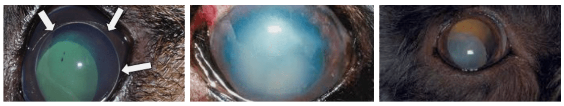
This often happens without you noticing and only an experienced ophthalmologist is likely to be able to pick up the earliest signs of lens luxation with special eye examination equipment. However, with time, the lens may become fully detached and will start to move freely. If the lens falls into the back of the eye, it may not be noticed for quite some time – but it is more likely that the lens will slip into the front of the eye – where it becomes stuck between cornea and iris. This is likely to cause acute pain and vision loss and you may notice that your dog’s eye is sore and blue.
Lenses that are trapped in the front of the eye are likely to cause increases in intraocular pressure - termed glaucoma – which can be blinding. Emergency treatment is usually required for lenses that have luxated into the front of the eye and your vet usually will try and recommend urgent referral upon diagnosis.
What happens if lens luxation is left un-treated?
If the lens is stuck in the front of the eye, it will almost always cause pain and vision loss – which with time becomes irreversible. Removal of the eye is then required to alleviate the pain. Lenses that are floating within the back of the eye tend to cause less acute damage but may also be harmful to the retina at the back of the eye with time.
Find out more about our ophthalmology referrals
How is lens luxation treated and what are the success chances?
There are many different treatment options for lens luxation – which is not always good indicator as this means that the perfect method for the treatment of a condition has not yet been found. Treatment options will also vary depending on whether your dog’s lens has already luxated or whether it is only unstable but still held in place. In general, the following options exist:
For lenses, that have luxated and are stuck in the front of the eye
The best treatment in this situation is to remove the lens from the front of the eye by surgery. This operation involves the use of the operating microscope and special microsurgical skills. The surgeon will usually have to make a long incision into the cornea to remove the luxated lens in its entirety. Possible complications of this operation include bleeding into the eye, retinal detachment or the development of high pressures (glaucoma). The chance, that a patient retains sight in the eye two years down the line after this operation is approximately 50%.
If surgery cannot be carried out (for example due to financial restraints), an attempt can be made to widen the pupil medically and then to push the lens into the back of the eye. This treatment usually requires a sedation. If the lens can be pushed back, then it is essential that the patient receives LIFE-LONG at least twice daily treatment with a drug that keeps the pupil small – which in turn ‘traps’ the lens in the back of the eye. This treatment approach is believed to be working in 8/10 patients – with an average chance of 50% that the eye will retain sight 12 months down the line.
If the lens cannot be pushed back and if surgery is not an option, then removal of the eye (enucleation) is the treatment of choice to provide pain relief.
For lenses that have luxated and are settled in the back of the eye
As for anterior luxated lenses, surgical removal is an option and the success chances are similar to that for lenses that are stuck in the front of the eye.
It may also be possible to keep these patients comfortable with varying degrees of vision with the use of eye drops to keep the pupil constricted. These drops MUST BE APPLIED TWICE DAILY WITHOUT FAIL as the luxated lens can otherwise come forward and become stuck if the pupil is dilated.
For lenses that are subluxated (unstable) but still in the correct place
Subluxated lenses can be removed via large incision surgery (as used for fully luxated lenses). The success chance with this operation is the same as for removal of fully luxated lenses – 50% of eyes having undergone this operation will remain sighted 2 years down the line.
Subluxated lenses can also be removed in a similar way as cataracts are removed – by a procedure called ‘phacoemulsification’.
In this operation, the wobbly lens is liquefied via a high-frequency ultrasound probe through a 2.8 mm incision into the eye. As the bag that has held the lens material will also be loose, it must be removed together with some of the gel that fills the back of the eye (a procedure called ‘vitrectomy’). This prophylactic small incision surgery has a high success rate and approximately 75% of patients are still sighted over 3 years after this operation. It is the least traumatic way to remove an unstable lens from an affected eye.
For patients where surgery is not ideal, we can attempt to maintain the lens in the back of the eye as it becomes more unstable. To this purpose, eye drops that keep the pupil small (so called ‘miotics’) are applied.
THESE DROPS MUST BE GIVEN TWICE DAILY WITHOUT FAIL.
Missing the drops might mean that the pupil could widen and that the lens, once it becomes fully loose, can then luxate into the front of the eye – where it will become acutely stuck and painful. The success chances of this so called ‘medical management’ of lens instability are unclear but some surgeons believe that they approach those of surgery – without the cost of a surgical procedure. The drugs used to keep the pupil small do not suit every patient and following the first application, the patient should be carefully observed for a couple of hours before long-term treatment is started.
Follow Us: